EYE FLOATERS
Some of you might have encountered tiny specks or strands floating in front of you but just can’t get rid of them? This might most probably be….FLOATERS!
You might find it especially common when looking
directly at a light background, or when feeling light-headed. The perception of
floaters is medically known as myodesopsia.
Floaters are those tiny spots, specks, flecks and
“cobwebs” that drift aimlessly around in your field of vision. Sometimes you
may momentarily confuse them with dust or tiny insects floating across in front
of the eye.

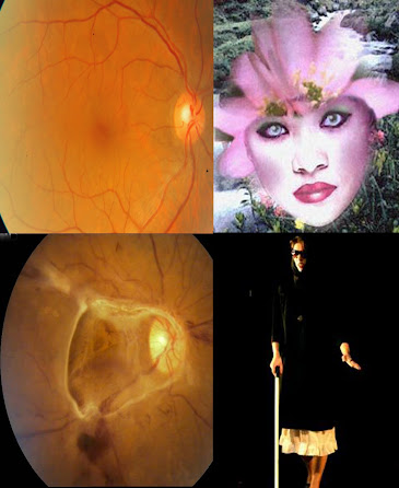
Fig. 1
Your eye is filled with a jelly like
substance called “vitreous Gel” (Fig.2). Light entering the eye, has to pass
through this gel to reach the retina. In childhood and adulthood, the vitreous
has a gel-like consistency. Any eye
condition in which the clarity of the vitreous humor is altered can produce the
symptom of eye floaters. As one gets older, changes normally begin within the
vitreous humor. The vitreous jelly naturally undergoes some liquefaction,
resulting in small pockets of more liquid vitreous lying within the firmer gel.
This is called vitreous syneresis. The boundary between each liquid pocket and
the gel may be noticeable to the person as one or more eye floaters. In
addition, it is normal for the collagen fibers that are within the vitreous to
become thickened and denser with age, resulting in eye floaters. Any person who
is over the age of 50 will have these changes within their eyes. However, the
degree of eye floaters produced by these typical changes will vary from person
to person.
Each
of these strands casts a small shadow onto the surface of the retina, and these
shadows may be perceived by you as eye floaters (Fig.3). As the eye moves from
side to side or up and down, these strands, deposits, or pockets also shift in
position within the eye, making the shadows move and appear to float or
undulate.
.jpg)
Fig. 2

Fig.3
You
may see the floaters as spots, straight and curved lines, strings, or "O"
or "C" shaped blobs. You may see it as a single floater while others
may think they see hundreds. The lines may be thick or thin, and they sometimes
appear to be branched. To most people, they appear grey and darker in color
than the background. The density of different eye floaters will vary within an
individual eye. Eye floaters may be more noticeable under certain lighting
conditions and be more apparent when looking at a bright sky. Floaters are
rarely seen in situations with reduced illumination.
Like
fingerprints, no two people have exactly identical patterns of eye floaters. If
you have eye floaters in both eyes, the pattern of the eye floaters in each eye
will be different. In any eye that has eye floaters, that pattern of eye
floaters may also change over time.
Usually floaters are harmless, though they are
annoying . Everyone experiences them from time to time and they cause no ill
effects.
Though your floaters are harmless, the following
changes in your floater should alarm you in having immediate medical attention.Floaters in
front of the eyes are normally clearly visible when looking into a light
background. However, if they start becoming visible in every background, if
they Suddenly Increase in Number and size, and are Accompanied
by Photopsia (perception of flashes of
light), it is vital that immediate medical advice is sought. This
could be an early sign of Retinal Detachment, which is a
serious blinding disorder. In such situations a prompt retinal evaluation is
needed, wherein they try to detect any retinal breaks or holes which may lead
to retinal detachment. If holes are detected and are treated with laser,
retinal detachment can be prevented.
But if you ignore these alarming symptoms and
wait till your vision falls, you might have developed retinal detachment. A
major surgery is needed to re attach the retina, which has poor prognosis.
Most
of the times, the floaters are due to normal aging changes of eye. Sometimes
they can be seen in children especially in high myopes ( with high minus glasses).
Floaters in these children can be harmless, if not associated with retinal
tears. Regular retinal evaluation is necessary.
Most eye floaters decrease in size and
darkness with time. Some of this is due to actual absorption of the floater
through the natural processes within the eye. Eye floaters may also shift in
position within the eye, resulting in less of a shadow effect. In addition, the
nerves within the brain adapt to and often become used to the presence of eye
floaters, ignoring them in a manner similar to the way a person only notices
the feeling of shoes on their feet when they think about it. Eye floaters
eventually tend to become less bothersome.
Eye
floaters cannot be removed with medication or other means. Most will fade over
time and become less annoying or noticeable.
There
are a large number of abnormalities in the eyes that may also cause the
symptoms of eye floaters. Any cellular material within the vitreous may cause
eye floaters. Red blood cells as a result of hemorrhage and white blood cells
as a result of inflammation are common types of cellular material causing eye
floaters. Hemorrhage into the vitreous may be a result of injury, diabetic
retinopathy, a retinal tear through a blood vessel, or eye surgery.
Inflammation in the vitreous may be caused by uveitis,
injury, infection, or eye surgery.

Fig. 4 Blood in vitreous
To
conclude, if you are above 50 years of age or if you are wearing minus glasses,
there are high chances of you seeing floaters. If you have floaters, do not
think and worry too much about them. But beware of warning signs and get
immediate retinal advice in such situation. Don’t take medications which are
falsely given to dissolve them. There is no medicine to dissolve them.

This is so interesting! I never knew eye floaters were actually little clumps of gel inside the eye. Thanks for the explanation!
ReplyDelete